選択した画像 sinus rhythm with bundle branch block 251169-Sinus rhythm with left bundle branch block
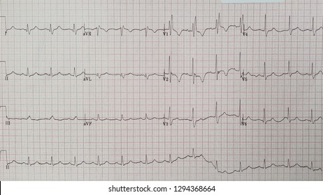
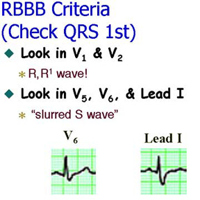
This occurs when a patient with fixed bundle branch block during sinus rhythm develops a reentrant tachycardia in the bundle branch system with a circuit with anterograde conduction over the non blocked bundle branch amd interventricular septal tissue, and retrograde conduction over the bundle branch which is anterogradely blocked during sinus rhythmSinus rhythm with a rate of 75 The QRS duration is wide at 146 ms There is a rsR' complex in lead V1 and a slurred Swave in lead I Bifascicular BlocksWide QRS complex (≥ 1 ms) Terminal Rwave in lead V1;
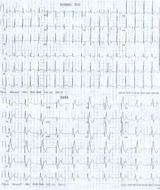
Pediatric Right Bundle Branch Block Background Pathophysiology Etiology
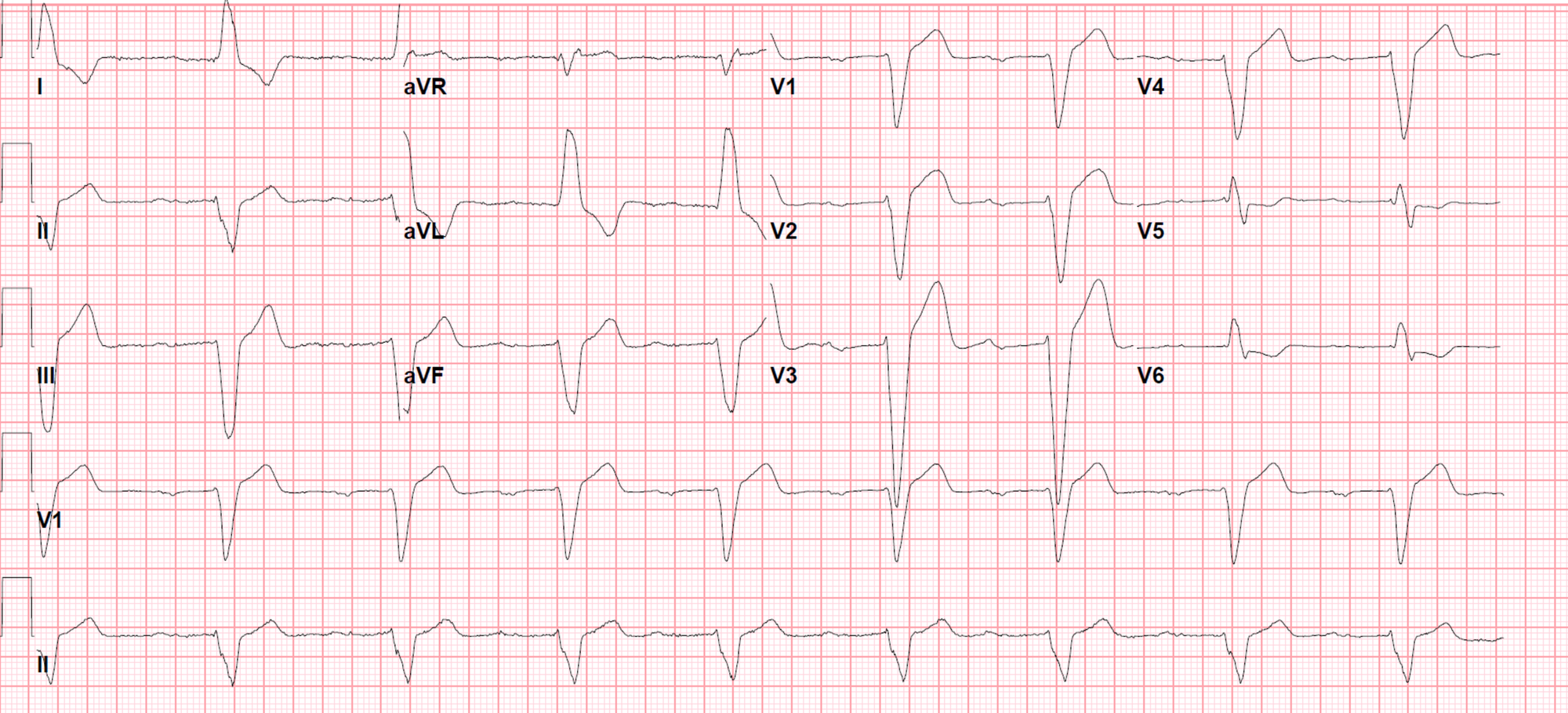
Sinus rhythm with left bundle branch block
Sinus rhythm with left bundle branch block-Rhythm analysis indicates a borderline normal sinus rhythm (NSR) Also present is a right bundle branch block (RBBB) Premature Atrial Complexes (PACs) exist in this encounter as well This encounter shows a borderline regular rhythm and heart rate, displaying P wavesSection 1 Supraventricular rhythms Normal sinus rhythm Normal sinus rhythm with a normal U wave Sinus arrhythmia (irregular sinus rhythm) Sinus tachycardia Sinus bradycardia Atrial bigeminy Atrial trigeminy Ectopic atrial rhythm Multifocal atrial tachycardia Atrial fibrillation Atrial fibrillation with rapid ventricular response Atrial fibrillation and bundle branch block Atrial


B26 Left Bundle Branch Block Lbbb Ecgs At St Emlyn S
There is sinus rhythm and Right Bundle Branch block There is a tall Rwave in V2 and V3 and slightly excessive ST depression in lead V2, with a biphasic wave What is it?1 doctor answer • 1 doctor weighed in Ecg Normal sinus rhythm, Incomplete right bundle branch block, borderline ecgRight bundle branch block comes from a problem with the heart's ability to conduct electrical signals It usually doesn't cause symptoms unless you have some other heart condition Your heart has 4 chambers The 2 upper chambers are called atria The 2 lower chambers are called ventricles In a healthy heart, the electrical signal for your heartbeat
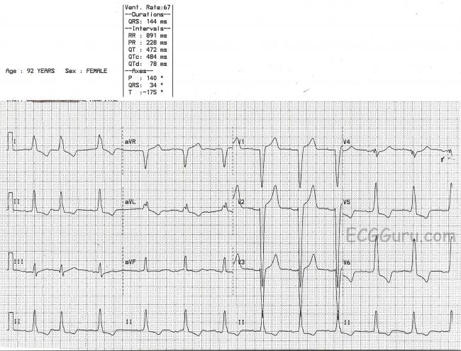
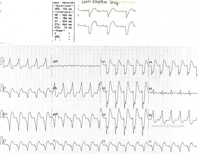
The electrical impulse in bundlebranch block originates in the sinus node, not in ventricular tissue, but a discussion of bundlebranch block is included in this rhythm group because of the location of the block within the ventricles and the wide QRS complex2613The ECG meets most of the criteria for left bundle branch block wide QRS, negative QRS in V1, positive QRS in Lead I and V6The axis is leftward, which is common in LBBB However, it is difficult to say for certain that this is a supraventricular rhythm Later, however, the patient's rate slowed (see top strip), revealing P waves When the rate slowed, the left bundle branch blockThe bottom ECG strip shows normal sinus rhythm with RBBB and acute ST elevation of >2mm Right bundle branch block is the result of a conduction delay or block within the right bundle branch Causes include right ventricular hypertrophy or strain, coronary artery disease, pulmonary embolism, WolffParkinsonWhite syndrome, myocarditis, and idiopathic causes
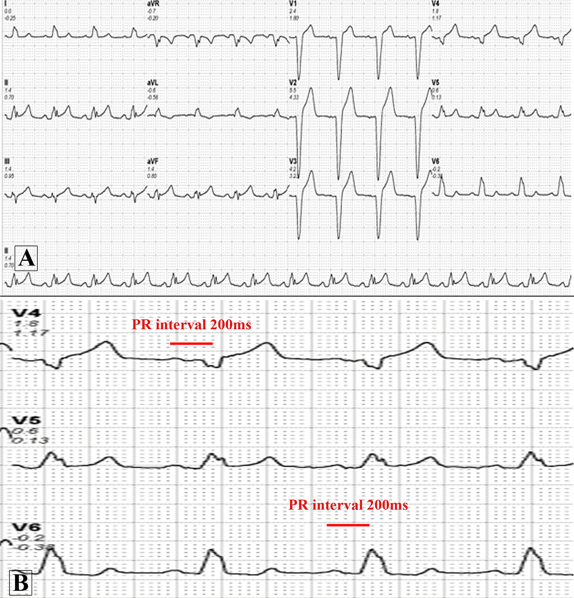
Occasionally, bundle branch blocks can arise intermittently, also making the diagnosis of ischemia challenging because the normally conducted beats now may show repolarization abnormalities, which is known as cardiac memory There are several proposed mechanisms of transient bundle branch blocks (1) a physiologic block during phase 3 and 4 of the action potential;Supraventricular tachycardia and sinus rhythm with contralateral bundle branch block patterns Han S(1), Miller JM(1), Das MK(1) Author information (1)Krannert Institute of Cardiology, Indiana University School of Medicine, Indianapolis, IN, USA A contralateral bundle branch block (BBB) aberration during tachycardia with a preexisting BBB strongly suggests the presence ofA 38yearold woman not on regular medications presented with a history of intermittent palpitations Her 12lead electrocardiogram (ECG) showed sinus rhythm with PR interval of 160 ms, QRS of 140 ms duration, and left bundle branch block (LBBB) patternAn echocardiogram demonstrated a structurally normal heart
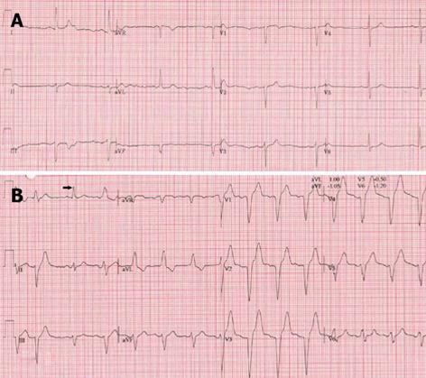


Exercise Induced Left Bundle Branch Block An Infrequent Phenomenon Report Of Two Cases


Intermittent Left Bundle Branch Block What Is The Mechanism
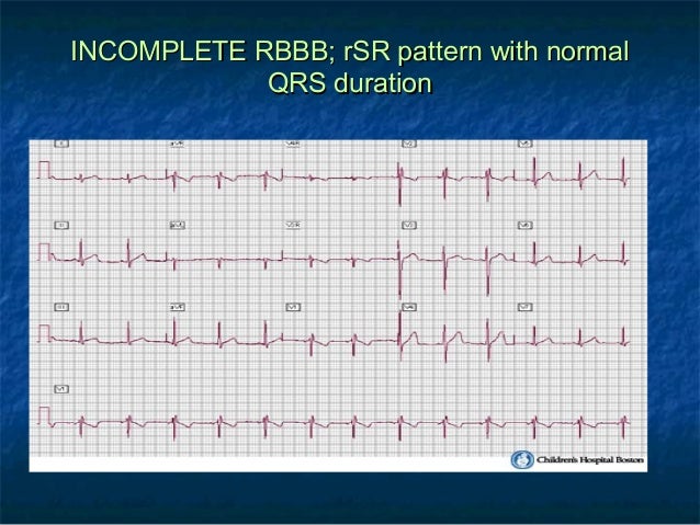
Wiring of heart Sinus rhythm means that the electrical activity that controls the contraction of the heart, starts in the sinus node Right bundle branch block means that the right bundle (part of the electrical wiring) does not conduct the impulse which changes the appearance of the ekg Right bundle branch block as opposed to left bundle branch block is usually benign0618A left bundle branch block refers to a disruption in the arrival of electrical impulses to the left ventricle of your heart We'll break down what this means and tell you about the kinds of0321Incomplete right bundle branch block is defined by QRS complex duration between 90 and 100 ms in children between 4 and 16 years of age, and between 86 and 90 ms in children less than 4 years of age 2 Incomplete RBBB may be diagnosed when the terminal rightward deflection is less than 40 ms but greater than or equal to ms 2


Electrocardiogram Sinus Rhythm Left Bundle Branch Block And Of Its Download Scientific Diagram


Q Tbn And9gcqts24zy8 O4ata1y1l95dt16bmfklbcgfih0gkrhxza2ulzs Y Usqp Cau
Left bundle branch block (LBBB) is the consequence of anatomical or functional dysfunction in the left bundle branch, causing the impulse to be blocked Depolarization of the left ventricle will be carried out by impulses spreading from the right ventricle Because the left bundle branch is dysfunctional, the impulse will spread (through the left ventricle) partly or entirely outside of theWhat is right bundle branch block?Click the link below to get FREE access to a massive library of helpful videos (not on Yout


Pin On Nursing Study Aids Mnemonics


A Normal Sinus Rhythm With Left Bundle Branch Block Download Scientific Diagram
Intermittent left bundle branch block in the hospital, her ECG revealed a very complex and interesting phenomenon Her baseline ECG (see Figure 1) showed sinus rhythm (SR), heart rate 87/min, left ventricular hypertrophy with repolarisation abnormalities and normal PR, QRS and QTc intervals Her baseline ECG transformed into sinus tachycardia with left bundle branch blockIs it posterior STEMI?0110Alternating Bundle Branch Block During Sinus Rhythm Show all authors Rakesh Sarkar 1 Rakesh Sarkar Department of Cardiac Electrophysiology and Pacing, Medanta—The Medicity, Gurugram, DelhiNCR, India See all articles by this author Search Google Scholar for this author, Kartikeya Bhargava 1 Kartikeya Bhargava Department of Cardiac Electrophysiology and


Left Bundle Branch Block Induced Cardiomyopathy In A Transplanted Heart Treated With His Bundle Pacing Jacc Case Reports


County Em Rhythm Nation Ecg Intermittent Left Bundle Branch Block
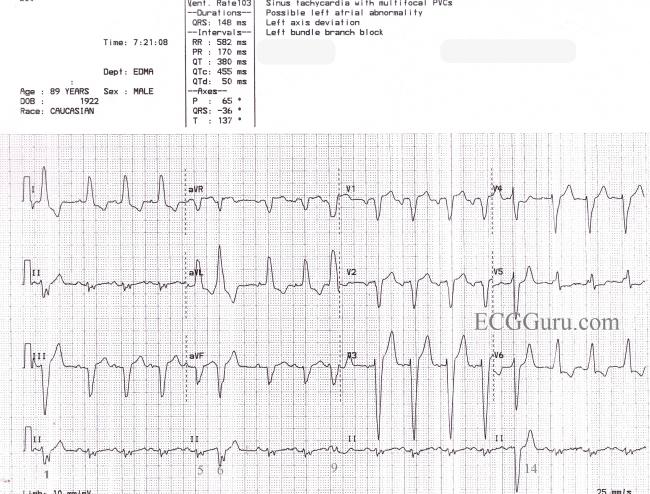
2304CRT should now be strongly considered in any patient with heart failure and bundle branch block As we have seen, bundle branch block causes the ventricles to beat sequentially (one after another) instead of simultaneously This discoordination of the normal pattern of ventricular contraction diminishes the efficiency of the heart beat In a person with a normalSlurred Swave in lead I;Sinus Rhythm With Left Bundle Branch Block, PVCs, and Fusion Beats This is a great ECG for teaching your students about some of the different causes of wide QRS This year old man has a sinus rhythm that is around 100 bpm, and his QRS is widened at 148 ms (148 sec)


Electrocardiography For Healthcare Professionals Ppt Download


Sinus Rhythm And Right Bundle Branch Block Configuration After The Download Scientific Diagram
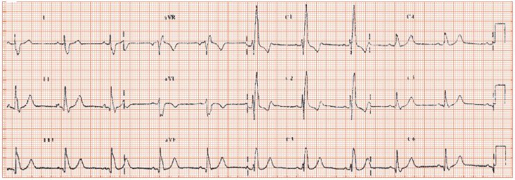
Footnote 12lead ECG shows sinus rhythm with right bundle branch block (RBBB) (wide rsR′ complex in lead V1 with a deep and wide S wave in lead V5 and V6) and left posterior fascicular block (LPFB) (with axis >1 Alternating RBBB and LBBB premature ventricular contractions 2 Alternating phase 3 block in the bundle branches 3 Alternating phase 4 block in the bundle branchesFinally, new right bundle branch block in patients experiencing dyspnea (particularly if acute) may indicate pulmonary embolism In the vast majority of cases, however, right bundle branch block is a benign finding with little if any impact of cardiovascular prognosis Young individuals rarely display right bundle branch block However, incomplete right bundle branch block occurs


Case B12 Junctional Rhythmn With Right Bundle Branch Block St Emlyn S Ecg Library St Emlyn S


B23 Right Bundle Branch Block Rbbb Ecgs At St Emlyn S
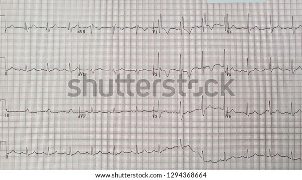
Sinus rhythm means your heart is beating at a normal and typical pace Marked sinus arrhythmia means that as you breathe, quite normally, your heart rate will increase as you inhale and decrease while you exhale, but in your case, it's varying a little more than typical This is astoundingly common, and normally means absolutely nothing0615C, ECG of an interfascicular reentry with right bundle branch block morphology and rightaxis deviation (type B) D, ECG during sinus rhythm of the patient with interfascicular reentry in , showing PR prolongation, right bundle branch block, and rightaxis deviation Note that QRS morphology in tachycardia is identical to that in sinus rhythmIn right bundle branch block, there is a problem with the right branch of the conducting system that sends the electrical signal to the right ventricle The electrical signal can't travel down this path the way it normally would The signal still gets to the right ventricle, but it is slowed down, compared to the left bundle Because of this, the right ventricle contracts a little later than it


Right Bundle Branch Block
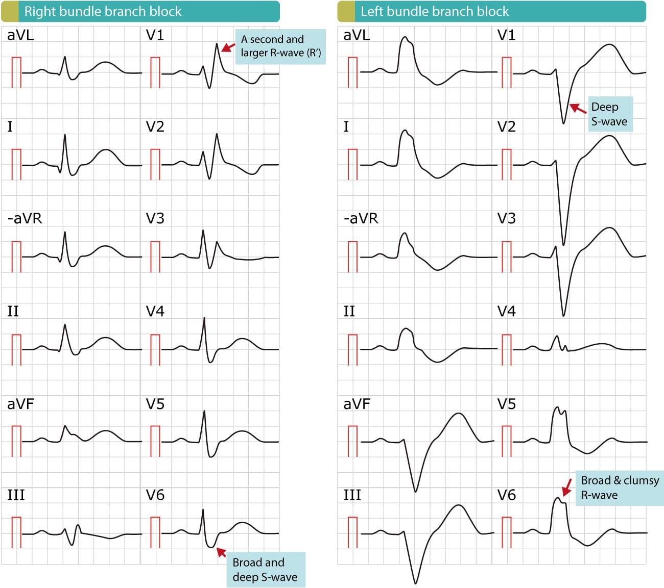


Right Bundle Branch Block Rbbb Ecg Criteria Definitions Causes Treatment Ecg Echo
Marked fibrosis of the sinus node was more frequent in patients with severe or moderate amyloid, but fibrosis of the bundle branch did not appear to be related to the amount of amyloid elsewhere in the heart Varying degrees of atrioventricular and bundle branch block were also present in six patients with no morphologic abnormalities of the conduction system Thus, conduction and rhythm0618This makes it harder for the heart to pump blood throughout your body Doctors call the resulting electrical pattern bundle branch block because the electrical impulse encounters a roadblock at theTwelvelead ECG showing sinus rhythm with alternating right and left bundlebranch block pattern of ventricular conduction Alternating RBBB and LBBB premature ventricular contractions Alternating phase 3 block in the bundle branches Alternating phase 4 block in the bundle branches Interpolated premature ventricular contractions with alternating bundlebranch
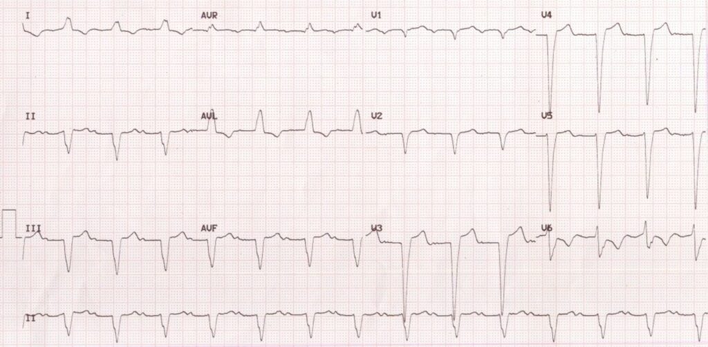


Lbbb With First Degree Av Block


Wide Qrs Complex Tachycardia With Alternating Right Bundle Branch Block Morphologies What Is The Mechanism Heartrhythm Case Reports
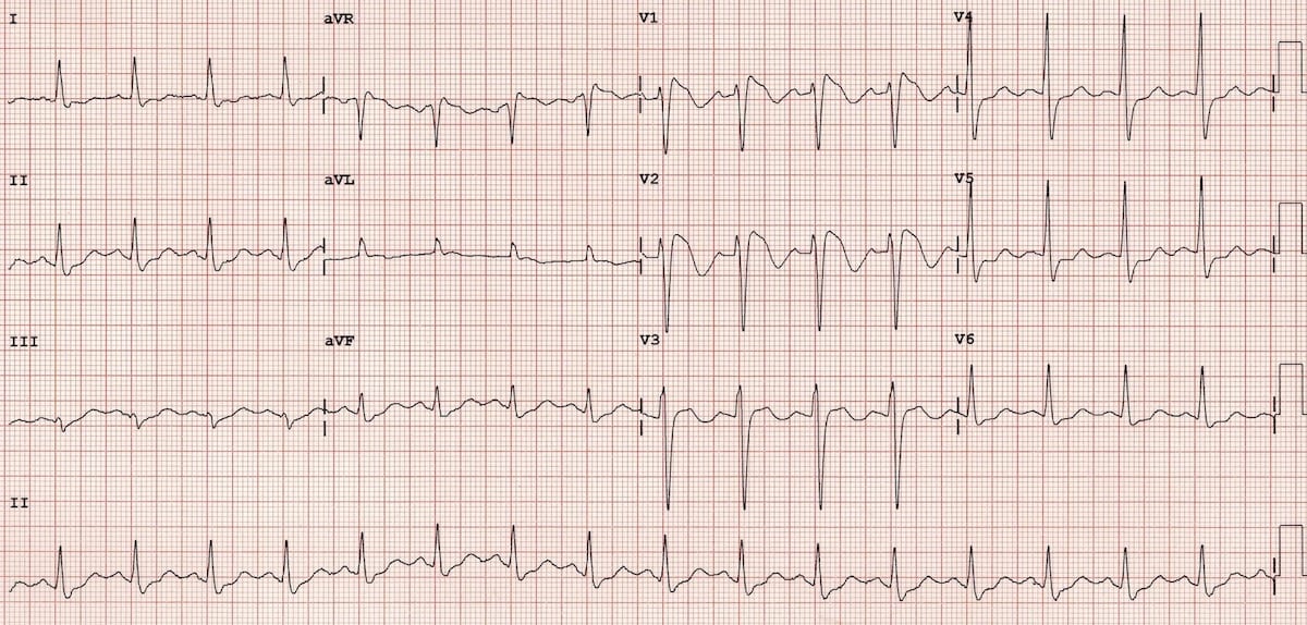
I took an EKG today and the results say sinus rhythm left atrial abnormality incomplete right bundle branch block What exactly could that mean?3003The bundle branch block can lead to a serious misdiagnosis during tachycardia Note the P wave following the ectopic pause (red highlight) confirming sinus tachycardia rather than ventricular tachycardia A "bradycardia dependent bundle branch block" is a rare ECG finding You may see a glimpse of it after a pauseSinus Rhythm There is an rSR' in V1, with wide Swaves in lateral leads (right bundle branch block, RBBB) Normally, RBBB has a bit of ST depression in V1V3 that is discordant (in the opposite direction of) the R'wave


Intro To Ekg Interpretation Bundle Branch Blocks Youtube
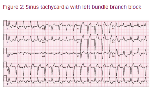


Intermittent Left Bundle Branch Block A Challenging Case Of Rare Electrocardiogram Phenomenon Touchcardio
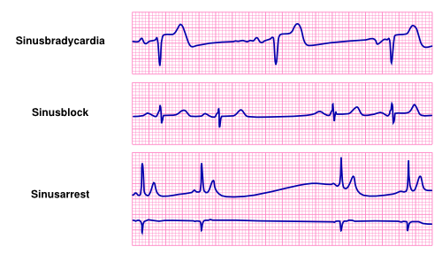
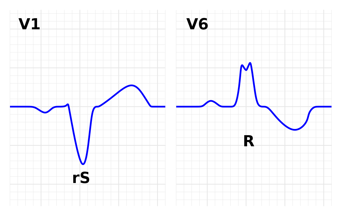
Left bundle branch block Inferior wall myocardial infarction Ventricular ectopic rhythms (eg, ventricular tachycardia) Congenital heart disease primum atrial septal defect, atrioventricular canal Hyperkalemia Emphysema Mechanical shift, such as with expiration or raised diaphragm pregnancy, ascites, abdominal tumor WolffParkinsonWhite syndrome, pacemakergenerated rhythmBundle branch blocks 43 Tachybrady syndrome 45 Treatments for heart blocks and slow If the sinus rhythm is slower than 60 bpm, it is called sinus bradycardia If the sinus rhythm is faster than 100 bpm, it is called sinus tachycardia ('Brady' means slow and 'tachy' means fast) The normal heart rate varies from minute to minute, depending on the demands on the heartLeft bundle branch block ECG characteristics of a typical LBBB showing wide QRS complexes with abnormal morphology in leads V1 and V6 Left bundle branch block (LBBB) is a cardiac conduction abnormality seen on the electrocardiogram (ECG) In this condition, activation of the left ventricle of the heart is delayed, which causes the left


Implications Of Left Bundle Branch Block In Patient Treatment American Journal Of Cardiology
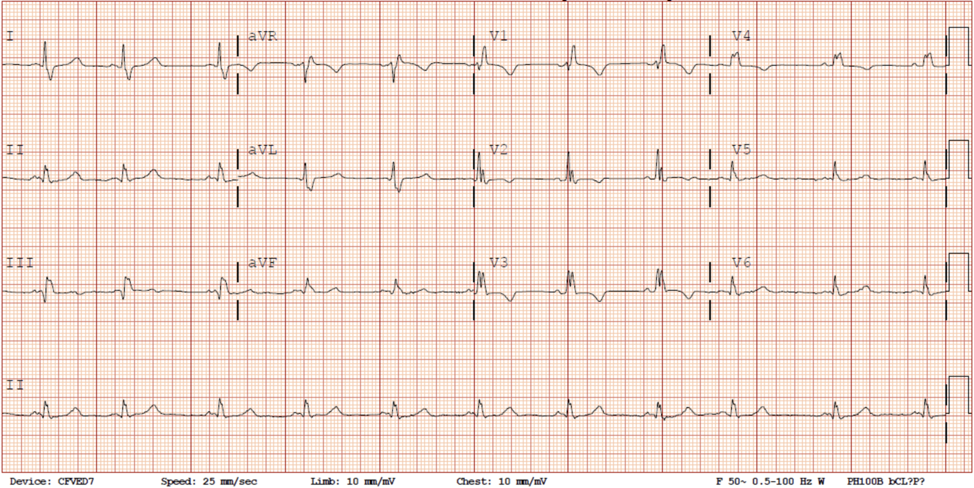


Cureus Transient Right Bundle Branch Block Resulting From A Blunt Cardiac Injury During A Motor Vehicle Accident
Figure 1 Holter Tracing of a Gentleman With Syncope Commentary The left half of Holter tracing in Figure 1 shows sinus rhythm withAnswer below Answer Here I have annotated the ECG The apex of the Twave is seen in V1 (inverted) If you draw a line down to V2, you see that the nadir of theRight bundle branch block with normal, In the other four with sinus rhythm the second degree block was localized distal to the BH deflection (HV) Most of the patients with RBBB (normal axis) and first degree AV block had a normal HV time with delay localized in the AV node (AH), indicating that this combination in the electrocardiogram does not necessarily mean bilateral bundle


Bilateral Bundle Branch Block During Sinus Rhythm A Left Bundle Branch Download Scientific Diagram
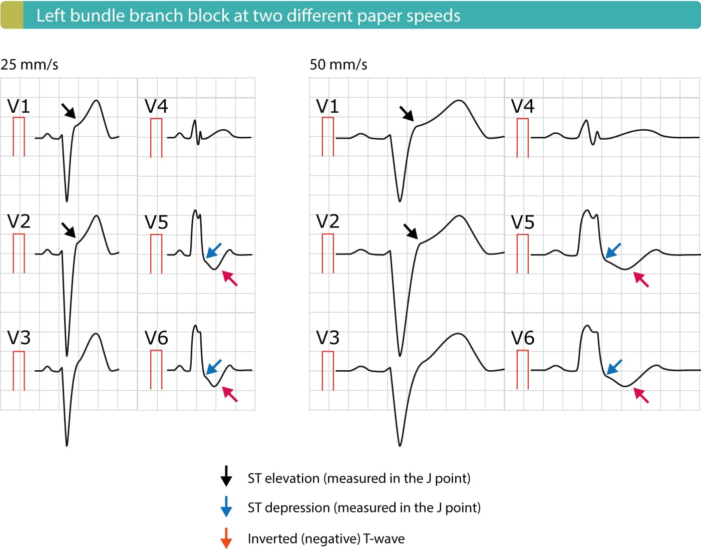


Left Bundle Branch Block Lbbb Ecg Criteria Causes Management Ecg Echo
Five and onehalf years later the block recurred and eight and onequarter years later it was replaced by sinus rhythm with the right bundle branch block—left anterior hemiblock pattern This patient demonstrates the lability of the right bundle branch block—left anterior hemiblock pattern in a surgical situation Even though complete heart block is replaced by sinus rhythm0707The rhythm strip shows sinus rhythm with two QRS configurations;(Figure 1) showed sinus rhythm with alternating pattern of right bundlebranch block (RBBB) and left bundlebranch block (LBBB) What is the mechanism of the alternating bundlebranch block pattern noted on the ECG?
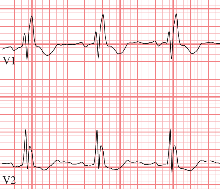


Tutorial Bundle Branch Blocks
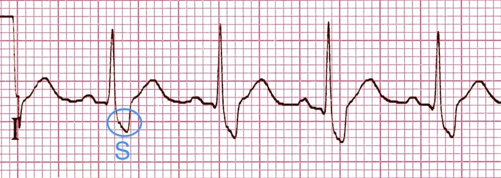


Right Bundle Branch Block Rbbb Litfl Ecg Library Diagnosis
What is a bundle branch block?Studying for a nursing school exam?ECG review of a Left Bundle Branch Block (LBBB) on a 12lead ECG with multiple ECG examples and images Criteria are included and Sgarbossa criteria, Cabrera's sign and Chapman's sign are100 degrees and no Q waves in I
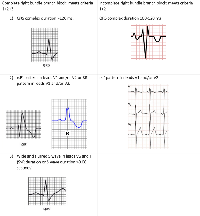


Diagnosis Of Right Bundle Branch Block A Concordance Study Bmc Family Practice Full Text


Electrophysiology Of The Heart Ecg Monitoring The Ecg
One narrow (yellow highlight) and one wide with a right bundle branch block configuration (red highlight) This is a bidirectional rhythm (or tachycardia if greater than 100 bpm) Let us look atECG on admission demonstrating normal sinus rhythm (62/minute), normal axis (15 degrees), PR interval of 016 seconds, and a normal QRS waveform Figure 2 ECG showing complete LBBB pattern on chest lead A CASE OF ALTERNATING BUNDLE BRANCH BLOCK 739 Vol 46 No 4 The patient had a history of diabetes that was diagnosed years previously for which she wasElectrocardiogram (EKG) revealed normal sinus rhythm (NSR) with a rate of 71 beats/min and left ventricular hypertrophy by voltage criteria An EKG on a previous admission showed NSR with a left bundle branch block pattern at a rate of 90 beats/min Carotid arteriograpby demonstrated left internal carotid stenosis with an ulcerated plaque The patient was premedicated with morphine
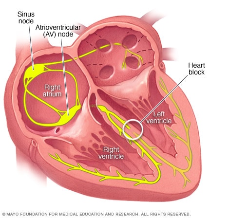


Bundle Branch Block Symptoms And Causes Mayo Clinic


Resolution Of Bundle Branch Block Circulation
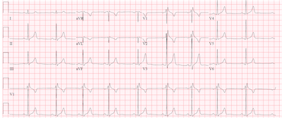
BACKGROUND Left bundle branch block (LBBB) is associated with atrial fibrillation (AF) and systolic heart failure, which can be treated with cardiac resynchronization therapy (CRT) that includes an implantable cardiac device (ICD) However, in some patients, LBBB may vary with heart rate, and during episodes of AF in LBBB, aberrant ventricular conduction, or wide QRS complexSinus rhythm with right bundle branch block (RBBB) and the echocardiogram was normal He underwent a 24hour Holter study, one of the tracings of which is shown in Figure 1 What does it reveal and what is the underlying mechanism of the finding?The ECG criteria for a trifascicular block on the 12lead ECG is reviewed including a right bundle branch block (RBBB), LAFB and 1st degree AV block


Heart Rhythm Guide
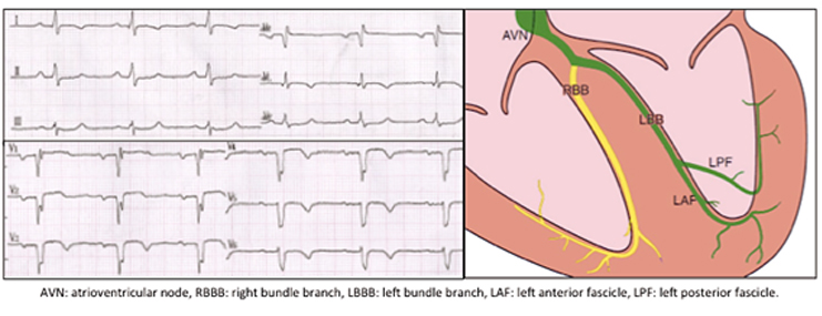


Tachycardia Dependent Bilateral Bundle Branch Block In Ischemic Heart Disease With Systolic Dysfunction Case Report And Review Of Prognostic Implications Medwave
Right Bundle Branch Block (RBBB) Rules for right bundle branch block Supraventricular rhythm;


Bundle Branch Block Wikipedia


Second Degree 2 1 Atrioventricular Block With Right Bundle Branch Block What Is The Mechanism Heart Rhythm


Ventricular Arrhythmias And Bundle Branch Block Flashcards Quizlet


Complete And Incomplete Left Bundle Branch Block Lbbb Ekg Ecg Ankara Kardiyoloji Kalp Hastaliklari Mete Alpaslan Doktorekg Com


1


Left Bundle Branch Block Ecg 4 Learntheheart Com
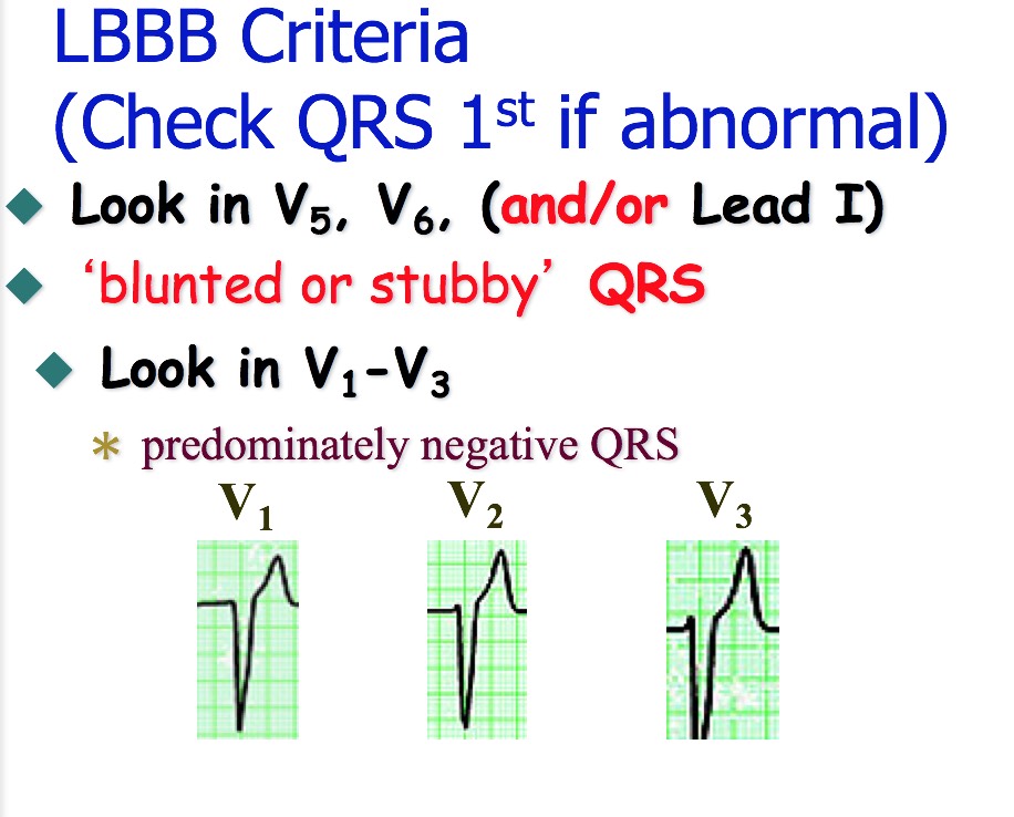


Bundle Branch Blocks


Ecg On Admission Sinus Rhythm Complete Right Bundle Branch Block Download Scientific Diagram


B26 Left Bundle Branch Block Lbbb Ecgs At St Emlyn S


Right Bundle Branch Block Part 1 Ems 12 Lead
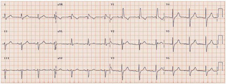


Right Bundle Branch Block Thoracic Key
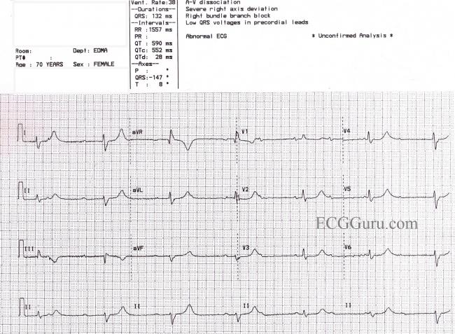


Third Degree Av Block And Junctional Escape Rhythm With Right Bundle Branch Block And Prolonged Qtc Interval Ecg Guru Instructor Resources


B Sinus Rhythm Left Axis Deviation Left Bundle Branch Block Download Scientific Diagram
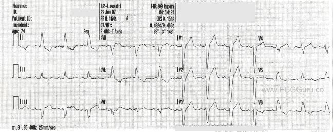


Left Bundle Branch Block Ecg Guru Instructor Resources


Bundle Branch Block Images Stock Photos Vectors Shutterstock


Right Bundle Branch Block Part 1 Ems 12 Lead
/right-bundle-branch-block-rbbb-1745785-FINAL-35a6f733fab44ef08240c21a675c12da.png)


Bundle Branch Block Overview And More


Ecg A Pictorial Primer


Category Bundle Branch Block


Complete Right Bundle Branch Block Of Unique Behavior What Is The Mechanism Akerstrom Cardiology Journal


Pediatric Right Bundle Branch Block Background Pathophysiology Etiology


Dr Smith S Ecg Blog Is This Left Bundle Branch Block Is There Stemi


Cureus A Curious Case Of Intermittent Left Bundle Branch Block Associated With Cough


Chest Pain Left Bundle Branch Block And Excessively Discordant St Segment Elevation Ems 12 Lead


Ekg S Internal Medicine University Of Nebraska Medical Center


Alternating Bundle Branch Block Circulation


Bundle Branch Block Wikipedia


Left Bundle Branch Block Ecg Guru Instructor Resources


Dr Smith S Ecg Blog New Left Bundle Branch Block Lbbb And Dyspnea


B Normal Sinus Rhythm Left Bundle Branch Block With Anterolateral T Download Scientific Diagram


Left Bundle Branch Block Lbbb Ecg Criteria Causes Management Ecg Echo


Pdf The Rate Dependent Bundle Branch Block Transition From Left Bundle Branch Block To Intraoperative Normal Sinus Rhythm Semantic Scholar


Right Bundle Branch Block And Presyncope In A 25 Year Old Man


Ecg Normal Sinus Rhythm With Left Bundle Branch Block And A Short Run Download Scientific Diagram


Q Tbn And9gcsztx Yiup6dgjcscknsguogls0mjbhalqbp1auz2 K1yzjb3ef Usqp Cau


Cureus Left Bundle Branch Block A Reversible Pernicious Effect Of Lacosamide


Bundle Branch Block Causes Symptoms Diagnosis Treatment


View Image


Sinus Rhythm With Left Bundle Branch Block Pvcs And Fusion Beats Ecg Guru Instructor Resources


Right Bundle Branch Block


Complete Right Bundle Branch Block Sinus Stock Photo Edit Now
/Bundle-Branch-Block-58960e843df78caebceafb79.jpg)


Bundle Branch Block Overview And More


Right Bundle Branch Block Wikipedia


Clinical Implications Of Electrocardiographic Bundle Branch Block In Primary Care Heart


Right Bundle Branch Block Thoracic Key


Complete Right Bundle Branch Block Of Unique Behavior What Is The Mechanism Akerstrom Cardiology Journal


Unusual Electrophysiologic Phenomena Thoracic Key


Dr Smith S Ecg Blog What Is Lurking Underneath This New Right Bundle Branch Block


Right Bundle Branch Block Rbbb Litfl Ecg Library Diagnosis


In Patients With Left Bundle Branch Block What S The Best Test For Cad Consult Qd


Right Bundle Branch Block Rbbb Litfl Ecg Library Diagnosis


1


Right Bundle Branch Block Ekg Examples Wikidoc


Wide Complex Tachycardia Left Bundle Branch Block With Subsequent Rhythm Strip Ecg Guru Instructor Resources


Bradycardia Textbook Of Cardiology


Resolution Of Bundle Branch Block Circulation


Ecg Sinus Rhythm 1st Degree Av Block Left Bundle Branch Block Download Scientific Diagram


Left Bundle Branch Block Wikipedia


Ecg After Intravenous Verapamil Showing Sinus Rhythm Incomplete Right Download Scientific Diagram


Ventricular Arrhythmias And Bundle Branch Block Flashcards Quizlet


A Normal Sinus Rhythm With Left Bundle Branch Block Download Scientific Diagram


Bundle Branch Blocks


Left Bundle Branch Block Wikiwand


His Bundle Pacing A New Frontier In The Treatment Of Heart Failure Aer Journal


Atypical Ischemic Repolarization In Right Bundle Branch Block Sciencedirect


Bundle Branch Block


Right Bundle Branch Block Rbbb Litfl Ecg Library Diagnosis


Right Bundle Branch Block Wikipedia


Prevention Of Cardiac Adverse Events Associated With The Use Of Drugs In Patients With Severe Mental Illness Case Report Revista Espanola De Cardiologia
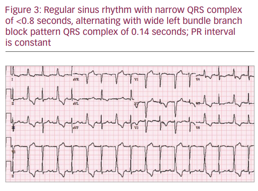


Intermittent Left Bundle Branch Block A Challenging Case Of Rare Electrocardiogram Phenomenon Touchcardio


Bundle Branch Block Wikipedia


B Normal Sinus Rhythm Left Bundle Branch Block With Anterolateral T Download Scientific Diagram


Intermittent Left Bundle Branch Block A Diagnostic Dilemma
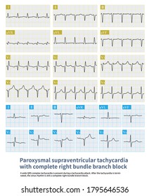


Bundle Branch Block Images Stock Photos Vectors Shutterstock


コメント
コメントを投稿